As I reflect on the progress in aesthetic medicine, particularly dermal filler treatments, it is evident that while these procedures offer fantastic opportunities for rejuvenation and enhancement, they come with challenges that need careful consideration. One recurring issue I’ve observed in practice is under-eye swelling, or malar oedema, an often distressing patient side effect.
Understanding the causes, management strategies, and evolving techniques for addressing these complications is essential for both practitioners and patients.
In this blog, I’ll explore the nuances of malar oedema,insights from the latest research, and practical solutions to improve patient outcomes.

What is Malar Oedema?
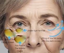
Malar oedema is swelling in the midface, typically under the eyes and extending to the cheek area. It can present as puffiness or fluid accumulation, disfiguring and persistent. Malar oedema is widespread after dermal filler treatments in the infraorbital hollow or tear trough region, where the lymphatic drainage system is delicate and prone to disruption.
The anatomy of the midface plays a crucial role here. The malar fat pad, a triangular structure in the cheek, and the malar septum, a fascial structure that acts as a barrier, can trap fluid when disrupted. This tr apping can be exacerbated by fillers that impede lymphatic flow, leading to prolonged swelling. The filler’s properties, injection depth, and patient factors such as pre-existing conditions or lifestylefurther influence the phenomenon.
How Dermal Fillers Contribute to Malar Oedema
While dermal fillers are designed to restore volume and contour, improper placement or overuse can lead to complications like malar oedema. This occurs due to:
- Compression of Lymphatic Structures: Fillers with high elasticity or excessive volume can physically compress lymphatic vessels, leading to fluid retention.
- Superficial Injection Techniques: Injecting fillers too close to the skin surface, above the malar septum, can exacerbate swelling.
- Patient Susceptibility: Conditions such as autoimmune disorders, viral illnesses, or dental infections can trigger inflammatory or hypersensitivity reactions in the filler-treated area.

Insights from Recent Research and Case Studies
The increasing prevalence of under-eye swelling and malar oedema in patients seeking filler treatments has been well-documented in recent literature. Here’s an overview of the key findings from the three papers I reviewed:
1. Funt’s Study on Avoiding Malar Oedema
Dr. David Funt’s article provides a detailed exploration of the anatomical causes of malar oedema and practical strategies to prevent it. His key findings include:
- Malar oedema is often caused by filler placement in the suborbicularis oculi fat (SOOF) and superficial malar fat compartments, where lymphatic drainage is already limited.
- Using preperiosteal injection techniques, where the filler is placed directly on the bone in small boluses, can help minimise lymphatic compression and reduce the risk of oedema.
- He emphasised the importance of limiting filler volume and using low-G’ (elasticity) fillers in the tear trough area to prevent complications.
- READ MORE HERE
2. The PHAREE Technique for Recurrent Eyelid Edema
This study introduced the PHAREE (Post-Hyaluronic Acid Recurrent Eyelid Edema) technique, which combines hyaluronidase injections with microneedling and small amounts of filler to treat persistent under-eye oedema.
- The researchers highlighted how this technique targets chronic oedema by breaking down excess filler and stimulating natural tissue healing.
- Early results from PHAREE show promising improvements in both aesthetics and patient satisfaction, though long-term outcomes remain to be studied.
3. Decates et al. on Under-Eye Swelling and Dental Health
A fascinating case report by Decates et al. examined a 32-year-old woman who developed unilateral facial swelling two weeks after receiving tear trough fillers.
- The swelling was ultimately traced back to apical periodontitis, a dental infection affecting the second bicuspid.
- This study underscores the importance of evaluating a patient’s overall health, including their dental status, before performing filler treatments. It also highlights the interconnectedness of facial anatomy and the need for multidisciplinary care.
- Read Here
Practical Recommendations for Patients and Practitioners
 As practitioners, we must approach dermal filler treatments carefully, particularly in high-risk areas like the tear trough. Based on the research and my clinical experience, here are some practical tips:
- For Practitioners:
- Always conduct a comprehensive patient evaluation, including medical and dental history.
- Use advanced injection techniques, such as the preperiosteal placement of small boluses, to minimise complications.
- Educate patients about potential side effects, including malar oedema, and set realistic expectations.
- For Patients:
- Be transparent about your medical history, including autoimmune conditions or recent illnesses.
- Understand that swelling is a possible side effect of fillers, and discuss strategies for prevention with your practitioner.
- Seek treatment from experienced professionals who prioritise safety and tailor treatments to your unique anatomy.
The Role of Emerging Techniques and Materials
Innovative approaches like the PHAREE technique and advancements in filler formulations are paving the way for safer and more effective treatments. However, as fillers continue to evolve, only time will tell how these solutions address long-term complications.
Despite these challenges, I firmly believe that dermal fillers remain a fantastic option for facial rejuvenation when used in small quantities and with skilled hands. With careful planning and patient-centred care, we can continue to achieve beautiful, natural results while minimising risks and remember with any aesthetic procedure risks and benefits are always part of consent – and it is important you understand these
Just My Thoughts
Tracey
read more here